Friday 27 May 2016

Coronary Artery Bypass Graft Surgery for Heart Patients - MyMedOpinion

Heart bypass surgery also called as coronary bypass surgery is done when coronary arteries in our heart are blocked or damaged. Coronary artery plays a vital role in supply of oxygenated blood to heart muscles, if blocked may result into restriction of blood supply flow and may lead to heart failure.


Heart Bypass surgery is commonly performed heart surgery in India which improves the blood flow to the heart with a new route or “bypass” around a section of clogged or diseased artery.
CABG (Coronary artery bypass graft)

Coronary artery bypass graft is most commonly performed and successful surgery for patients who have severe coronary heart disease (CHD). During this surgery, a vein or artery from another part of the body such as leg, arm, chest or abdomen is used to create a new path for blood to flow to the heart, bypassing the blocked artery. It restores and improves the pumping ability and blood flow to the heart muscle and thus prevents from heart attacks.

When it is recommended ?
Presence and severity of CHD symptomsSevere chest pain caused by narrowing of several of the arteries that supply your heart muscle, leaving the muscle receiving inadequate bloodA medical condition when more than one diseased coronary artery and left main coronary artery is severely blocked or narrowedA medical condition when heart’s main pumping chamber, the left ventricle isn’t working properlyA medical condition of failure or attempted angioplasty or stent placement and restenosiscall-back-buttonAdvanced & latest Minimal Invasive Heart Surgery


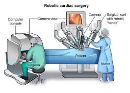
Minimal invasive Heart-SurgeryWith the advancement in medical technology, our surgeons are commonly providing relief to patients suffering with coronary artery disease through minimally-invasive procedure called trans-abdominal Coronary Artery Bypass Graft (CABG). it is performed while your heart is still beating that is without stopping the heart.

In traditional CABG surgery, surgeons were putting the patient on a heart-lung machine to stop the heart during the procedure. Now with this unique and most advanced approach, surgery is performed without stopping the heart. This surgical approach has been proven to be as safe and most successful with benefiting the patient into quicker recovery and to resume an active lifestyle.

Advantages of Minimally Invasive Bypass Surgery

The benefits of minimally invasive bypass surgery includes:
Reduced post-operative pain and less blood lossFewer traumas due to the elimination of the heart-lung machineCosmetically beneficial since smaller incision, thus a smaller scarQuicker recovery and to resume an active lifestyle

Thursday 28 April 2016

Coronary bypass surgery performed in world class hospitals in India is a procedure to allow blood to flow to your heart muscle despite blocked arteries. Coronary bypass surgery uses a healthy blood vessel taken from your leg, arm, chest or abdomen and connects it to the other arteries in your heart so that blood is bypassed around the diseased or blocked area. After a coronary bypass surgery, normal blood flow is restored. Coronary bypass surgery is just one option to treat heart disease.
Just like all the other organs in your body, your heart needs blood and oxygen to do its job. Coronary arteries snake across the surface of your heart, delivering a constant supply of blood and oxygen to the heart muscle. When one or more of these arteries become narrowed or blocked, blood and oxygen are reduced and heart muscle is damaged. Coronary bypass surgery can minimize this damage.
Today Cardiology treatment in India has come up as a suitable option in order to get rid of any of the heart defects as the cost in India of any of the treatments is the best and that too at rates which are absolutely affordable. Because of these benefits of choosing in India, any of the treatments, many foreigners have come down here in order to solve their trouble of heart diseases.


Who Should Consider Coronary Artery Bypass Graft Surgery?
• People diagnosed with arterial blockage or heart damage are recommended with the Coronary Artery Bypass Graft Surgery.
• People suffering from severe chest pain or angina due to the arterial blockage are recommended with the Coronary Artery Bypass Graft Surgery.
• People suffering from complicated conditions such as diabetes & high blood pressure are recommended the Coronary Artery Bypass Graft Surgery to reduce the risk of heart attack.

Procedure for Coronary Bypass Surgery
During a Coronary Artery Bypass Graft Surgery (CABG), the blood flow is re-routed around the clogged artery by detaching a long segment of an artery from the chest wall, arms or leg veins. Thereafter, the new artery is grafted to the clogged area of the coronary artery. Through the newly attached channel, blood gets unhindered route to flow to the heart muscles. This procedure is known as Coronary Artery Bypass Surgery. Depending upon the number of blocked coronary arteries, a patient may undergo more than one bypass graft.


What to Expect After Coronary Artery Bypass Graft Surgery?
After surgery, there will be a short stay (1 to 2 days if there are no complications) in the intensive care unit (ICU). In the ICU, you will likely have:

• Continuous monitoring of his or her heart activity.
• A tube to temporarily help with breathing.
• A stomach tube, to remove stomach secretions until the person starts eating again.
• A tube (catheter) to drain the bladder and measure urine output.
• Tubes connected to veins in the arms (intravenous, or IV, lines) through which fluids, nutrition, and medicine can be given.
• An arterial line to measure blood pressure
• Chest tubes, to drain the chest cavity of fluid and blood (which is temporary and normal) after surgery.


Benefits of Coronary Artery Bypass Graft Surgery
Some of the potential benefits of Coronary Bypass Heart Surgery (CABG) include :

• Lower risk of stroke
• Lower death rate
• Less need for transfusion
• Less heart rhythm problems
• Less injury to the heart

Monday 25 April 2016

Deposition of fatty substances in the coronary arteries (arteries supplying blood to the heart), develops a blockage. This condition is called as ‘coronary artery disease’ (CAD). This blockage creates a problem in the free flow of the blood to your heart muscle. This situation might lead to a heart attack. Angioplasty is a widely performed procedure that is used to treat heart problems caused by blocked or narrowed coronary arteries (blood vessels that supply blood to the heart muscle). This procedure is performed highly successfully by interventional cardiologists at heart hospitals in India. The coronary angioplasty, which normally takes between one to two hours, aims to remove or reduce blockages in the coronary arteries and restore blood flow to the heart muscle. The coronary angioplasty procedure is performed in a Cardiac Catheterisation Lab . All cardiac speciality hospitals in India as well as most of multi-speciality hospitals in India are equipped with Cath Labs


Procedure
Angioplasty procedure involves insertion of catheter in your groin and hence before the angioplasty procedure you would be given a mild sedative and some pain killers to ensure that you remain relaxed during the procedure. Your doctor will insert a catheter into an artery in your groin and gently manoeuvre it to the heart. This catheter has a balloon at the tip and once the catheter reaches the site of the blockage, the balloon on the end is inflated. This opens the blockage and restores the proper flow of the blood to the heart. Often the device which is called a stent is also placed at the site of blockage in order to keep the artery open.
A stent is a small, mesh like metallic device, which when placed at the site of blockage acts as a support to keep the artery open thus maintaining the blood flow. Two main varieties of stents are used. The first is a non medicated or bare stents and the second is medicated or drug eluting stents. Both the stents serve the function of supporting the open artery. The medicated stent in addition releases drug locally at the treated site to reduce the chances of recurrence of blockage at the same site.


Advanced Interventional Cardiology Procedures for Treatment of Heart Diseases in India | Best Heart Surgery Hospital India
Balloon Angioplasty - Balloon Angioplasty is a procedure in which a narrowed heart valve is stretched open in a way that does not require open heart surgery.It is a procedure in which a thin tube (catheter) that has a small deflated balloon at the tip is inserted through the skin in the groin area into a blood vessel, and then is threaded up to the opening of the narrowed heart valve. The balloon is inflated, which stretches the valve open. This procedure cures many valve obstructions. It is also called balloon enlargement of a narrowed heart valve.


Stents - Approximately 70% of angioplasty procedures also involve stenting, which is the insertion of a small metal cylinder called a stent into a blood vessel. In this procedure, a collapsed stent is placed over the balloon at the tip of the catheter. When the balloon inflates, the stent pops open and reinforces the artery walls. The balloon and catheter are then withdrawn and the stent inside permanently. In a few weeks, tissue from the artery lining grows over the stent.There are two types of stents. Bare-metal stents are plain, untreated metal cylinders. Drug-eluting stents (also called drug-coated stents) are coated with medication before they are placed in the artery.


Rotablation (Percutaneous Transluminal Rotational Atherectomy or PTRA) - A special catheter, with an acorn-shaped, diamond-coated tip, is guided to the point of narrowing in the coronary artery. The tip spins around at a high speed and grinds away the plaque on the arterial walls. This process is repeated as needed to treat the blockage and improve blood flow. The microscopic particles are washed safely away in your blood stream and filtered out by your liver and spleen.


Drug-Eluting Stents - A drug-eluting stent (DES) is a coronary stent (a scaffold) placed into narrowed, diseased coronary artery. It slowly releases a drug to block cell proliferation. This prevents fibrosis that, together with clots (thrombus), could otherwise block the stented artery, a process called restenosis. The stent is usually placed within the coronary artery by an Interventional cardiologist during an angioplasty procedure.


Angioplasty vs. Bypass Surgery
It is not easy for a patient to make a choice between angioplasty and bypass surgery on his own, which happens due to various factors like the condition of the disease in an individual. The doctor also checks the symptoms of the patient along with his overall heart function and co-existing medical conditions.

In case the narrowing in the arteries is serious about reducing the blood flow in the heart of the patient then angioplasty is recommended. But if the patient has various blockages, bypass surgery is certainly better.

Friday 15 April 2016

Affordable Heart Surgery at Best Hospitals in India

Best Heart Surgery Hospitals in India 


Your Heart pumps blood through the circulatory system to all parts of the body and hence it is known as the blood-pump of the body. The blood thus circulated carries food and oxygen to the cells (so that they can function properly) and carries away carbon dioxide and waste materials.
A very large number of people suffer from heart disease and millions of them die each year worldwide. There are many types of heart diseases that affect your heart. The various heart diseases include diseases of your blood vessels, such as coronary artery disease, heart rhythm problems heart infections and heart problems that you may have at the time of birth.
Sometimes the term "heart disease" is often used interchangeably with "cardiovascular disease." Cardiovascular disease is caused by disorders of the heart and blood vessels, and includes coronary heart disease (CHD) cerebrovascular disease (stroke), raised blood pressure, peripheral artery disease, rheumatic heart disease, congenital heart disease and heart failure. Cardiovascular diseases are the number one cause of death globally and more people die annually from cardiovascular diseases than from any other cause. Some of the common causes of cardiovascular disease are tobacco and excess alcohol use, physical inactivity, stress and an unhealthy diet.

Common Cardiovascular Diseases

  • CoronaryHeart Disease (CHD): This is also known as Ischaemic Heart Disease (IHD) or Coronary Artery Disease (CAD)and is the most common type of cardiovascular disease in many countries around the world. In simple, Coronary Heart Disease refers to problems with the circulation of blood to the heart muscle. The circulation of the blood may be affected due to one or more blockages in the coronary arteries. Coronary arteries are the vessels that deliver oxygen-rich blood to heart muscle. A partial blockage in the coronary arteries may cause symptoms such as chest pain (angina) and shortness of breath. A complete blockage of an artery may causedamage to the heart muscle tissuesand may lead to a heart attack. In some people there may be multiple blockages.
  • Cerebrovascular Disease (Stroke): Just like heart muscle, your brain also needs a constant supply of oxygen and nutrients to function properly. For this you should have an interrupted circulation of blood to the brain. In some people the circulation of blood in the blood vessels of the brain might get disturbed. Because of this the cells may start dyeing due to the lack of oxygen rich blood. Such a situation may cause a stroke. A stroke is an emergency medical condition since it may cause serious brain damage and possibly death if the treatment is delayed. The sooner you get the treatment for stroke lesser the damage is likely to occur.
  • Peripheral Vascular Disease: In Peripheral Vascular Diseasethe blood circulation is affected due to blockage in the arteries to your limbs, primarily in the legs. If you have Peripheral Vascular Disease then you may have pain in the calves, thighs or hips especially when walking, running or climbing stairs.You may feel cramps, a dull pain or a sensation of heaviness in the muscles of your legs.Peripheral vascular disease is also known as Peripheral arterial disease.
  • Cardiomyopathy: Cardiomyopathy refers to the diseases of the heart muscle where the heart muscle becomes enlarged, thick or rigid. If the cardiomyopathy worsens then the heart becomes weaker. In such a situation, the heart may not be able to properly pump blood through the body and maintain a normal electrical rhythm. This can lead to weakening of the heart orheart failure which in turnmay causefluid to build up in the lungs, ankles, feet, legs, or abdomen and in some cases it might also cause heart valve problems.There are different causes, signs and symptoms, and treatments available.
  • Valvular Heart Disease: Valvular Heart Diseaseis the disease of the valves of the heart. Your heart is a pump that pumps blood through the circulatory system in your body. The blood carries all the vital things that help your body function.The direction of blood flow is controlled by a set of four valves. Each valve opens and closes once per heartbeat. The valvesplay a major role in helping the blood to flow into the main arteries in one direction and preventingthe regurgitation of blood in reverse direction. In case of Valvular Heart Disease, the heart valves become less effective in controlling the flow of blood. The valves may fail to close fully or leak or the outlet of the valve may become narrowed. The problems with the valves maylead to the reduction of the blood flow and an increase in the pressure on heart wall.It may also cause back pressure in the lungs or the peripheral tissues. Valvular Heart Disease cause significant reduction of effective blood circulation causing congestion of the lungs and peripheral tissues. It may also lead poor oxygen supply to the vital organs.
  • Rheumatic Heart Disease: Rheumatic Heart Diseases are the heart disorders that can occur as a result of rheumatic fever. Rheumatic Heart Disease may cause damage to the heart valves. This may lead to a heart valve disorder.Rheumatic Heart Disease may affect heart valves during childhood and the actual heart problems may develop or may be seen in later years. In most of the cases the affected valves have to be replaced by a valve replacement surgery.
  • Congenital Heart Disease: Congenital Heart Disease is the disease that arises due to the defect/s in the structure of the heart that is present at birth. These defects can be as simple as a small hole in one of the inner walls of the heart or they can be very complex which may seriously affect the flow of blood in heart and lungs. Some congenital heart problems may need surgical intervention and in some cases more than one operation may be required.

Tuesday 5 April 2016

Ventricular Septal defect is a hole (defect) in the wall that separates the lower chambers of the heart. The lower chambers of the heart are called the ventricles. The wall between them is called the ventricular septum. In the normal heart, the septum prevents blood from flowing directly from one ventricle to the other. In a heart with a VSD, blood can flow directly between the two ventricles. VSD is a congenital (present at birth) heart defect. As the fetus is growing, something occurs to affect heart development during the first 8 weeks of pregnancy, resulting in a VSD.


Some terms to remember re: Congenital Heart Diseases
Atrioventricular Septal Defect (AVSD) - This includes an Atrial Septal Defect,Ventricular Septal Defect and abnormal development of the atrioventricular valves (tricuspid and mitral). This causes blood to flow abnormally inside the heart. An AVSD is also known as an atrioventricular canal defect.
Atrial Septal Defect (ASD) - This is a hole in the wall that separates the upper chambers (atria) of the heart. This causes blood to leak from one atrium to the other.

Patent Ductus Arteriosus (PDA) - In this condition there is a persistent connection between the aorta and the pulmonary artery. This connection is called the ductus arteriosus and is normally present before birth. In most babies, the vessel closes within a few hours to days after birth. In some children, this vessel fails to close.

• Problems with the Heart Valves
  1. o Aortic Valve Stenosis is a defect of the aortic valve in the heart that often causes it to open incompletely. This reduces blood flow to the body.

  2. o Pulmonary Valve Atresia is a defect where a solid sheet of tissue forms instead of the pulmonary valve. This prevents oxygen-poor blood in the right side of the heart from traveling normally to the lungs to pick up oxygen.

  3. o Pulmonary Valve Stenosis is a narrowing of the pulmonary valve. This slows the flow of blood from the right side of the heart to the lungs. The heart must pump harder to push blood through the smaller opening to the lungs where the blood picks up oxygen.

  4. o Tricuspid Atresia is a defect where a solid sheet of tissue forms instead of the tricuspid valve. The tricuspid valve is between the right atrium and right ventricle of the heart. Without the tricuspid valve, oxygen-poor or blue blood entering the right atrium cannot travel normally to the lungs to pick up oxygen.

  5. o Ebstein's Anomaly is a defect where the tricuspid valve is both displaced and abnormally formed. The valve is leaky and allows blue blood to flow back into the right atrium instead of to the lungs to pick up oxygen.
• Transposition of the Great Vessels - This is a defect where the location of the aorta and pulmonary artery coming off the heart is switched. The aorta comes off the right ventricle instead of the left ventricle. The pulmonary artery arises from the left ventricle instead of the right ventricle. Therefore, blood without oxygen is continually pumped to the body, instead of blood with oxygen.

• Tetrology of Fallot - is a combination of four defects:
  1. o Pulmonary valve stenosis (narrowing)

  2. o Ventricular Septal Defect

  3. o Overriding of aorta where the aorta is positioned between the left and right ventricles, over the Ventricular Septal Defect.

  4. o Right ventricular hypertrophy is the thickening of the right ventricle.
• Truncus Arteriosus - This is a defect of the aorta and pulmonary artery. The aorta and pulmonary artery do not form as separate arteries. Instead, a large artery, called the truncus, comes from the heart. As the truncus leaves the heart, it may branch into arteries that carry blood to the body and to the lungs.

• Coarctation of the Aorta - In this condition there is a narrowing of the aorta. It slows or blocks the flow of blood from the heart to the body.

What are the types of Ventricular Septal Defects?
Ventricular Septal Defect can be divided in to 3 types:
• Size of the Ventricular Septal Defect
  1. o Small Ventricular Septal Defect

  2. o Moderate (or medium-sized) Ventricular Septal Defect

  3. o Large Ventricular Septal Defect
• Location of the Ventricular Septal Defect
  1. o Membranous Ventricular Septal Defect are located near the heart valves.

  2. o Muscular Ventricular Septal Defect are found in the lower part of the septum

  3. o Inlet Ventricular Septal Defect are located close to where blood enters the heart.

  4. o Outlet Ventricular Septal Defect are found in the part of the ventricle where the blood leaves the heart
• Number of defects

• Presence or absence of a ventricular septal aneurysm

What Cause Ventricular Septal Defect?
Heredity may play a role. Parents who have congenital heart defects are more likely to have a child with VSD than parents who do not have congenital heart defects. In some cases, VSD may be due to a defect in one or more genes or to chromosomal abnormalities.


What are the Symptoms of Ventricular Septal Defect?
The following are the most common symptoms of VSD. However, each patient may experience symptoms differently. Symptoms may include:
  • fatigue
  • sweating
  • rapid breathing
  • heavy breathing
  • congested breathing
  • disinterest in feeding, or tiring while feeding
  • poor weight gain
The symptoms of VSD may resemble other medical conditions or heart problems.


How is Ventricular Septal Defect Surgery Performed?
The surgery to close a Ventricular septal defect is done under general anesthesia so that your child will be asleep and feel no pain. The surgeon makes a cut down the center of the chest to reach the VSD.


What is the outcome of surgery for Ventricular Septal Defect?
In 99% of cases, there are successful results with no complications following surgical closure of Ventricular Septal Defect in children as they have a remarkable ability to heal and recover quickly. There is usually no residual leakage. After the Open Heart Surgery to repair Ventricular Septal Defect, there might be some pain and discomfort which will soon subside. Your child will be able to participate in all the normal activities pretty soon leaving all the memories of pain and discomfort behind.

Robotic Heart Surgery at World Class Hospital in India

Robotic surgery for cardiac is a form of heart surgery performed through tiny incisions in the chest. The procedure is sometimes called da Vinci surgery because that is the name of the manufacturer of the robot often used for this procedure Robotic surgeries have been used for a number of different heart-related procedures, including mitral valve repair, coronary artery bypass, heart defect repair, and tumor removal.

Robotic Heart Surgery in India is the latest and most advanced technique in the world to perform heart surgery with greatest precision Robot Assisted Heart Procedures are done to treat a variety of conditions:
• Mitral valve repair—surgery to repair problems with the valve between the upper and lower chambers on the left side of the heart
• Coronary artery bypass grafting (CABG)—surgery that creates a path around blocked heart vessels so that blood can reach the heart muscle. The bypass is done using blood vessels from other parts of the body to
Atrial septal defect repair—surgery to close a hole in the wall between the upper two chambers of the heart
• Biventricular pacemaker lead placement for heart failure—surgery to place leads (wires) on the inner surface of the heart’s ventricles. The leads are attached to a biventricular pacemaker to help the heart beat in a regular rhythm.

Da Vinci Robotic system
The da Vinci robot is a highly sophisticated tool that enables the surgeon to perform the procedure with greater precision and control than is possible in traditional hands-on surgery. In robotically assisted surgeries, the chest cavity is accessed through dime-size "keyhole" incisions.
The surgeon sits in a console equipped with controls that direct robotic arms to perform the surgery. The da Vinci's robotic arms are essentially an extension of the surgeon's hands. A tiny camera attached to a robotic arm gives the surgeon a very detailed, three-dimensional view of the operating space inside the chest. The robotic arms are very agile, providing the surgeon a greater range of motion than is possible with hand-manipulated moves in standard procedures


Benefits of Robotic Heart Surgery
Minimally invasive surgery offers several advantages compared to open-chest procedures, including:

• Faster return to normal activities. Rather than waiting several weeks to heal, patients can return to work or other activities much more quickly--usually within 10 days.
• Shorter hospital stay. Time spent in the hospital can sometimes be reduced by as much at 50 percent, compared to open procedures.
• No splitting of the breastbone ("cracking the chest"). Keeping the breastbone intact reduces the chance for post-surgical complications and infection.
• Smaller incisions. Depending upon the case, the operation may be performed through four to five fingertip-size incisions or through a 3- to 5-inch incision at the side of the chest. Traditional open-heart procedures require a longer incision down the center of the chest.
• Significantly less pain. Decreased damage to tissue and muscle results in less pain. Tylenol or aspirin are often enough to manage pain after some procedures.
• Elimination of the heart-lung bypass machine, in most cases. Avoiding the bypass machine decreases the risks for neurological complications and stroke.
• Minimal blood loss and less need for transfusion.
• Little scarring. Instead of a long chest scar, only a few tiny scars or a short, 3- to 5-inch scar remains.

Tuesday 9 February 2016

Surgery for Atrial Septal Defect (ASD) in India | Best Heart Surgery Hospital India

What is Atrial Septal Defect (ASD)
Atrial Septal Asd Surgery, Atrial Septal Asd India
An atrial septal defect (ASD) is a hole in the wall between the two upper chambers of your heart. The condition is present from birth (congenital). Smaller atrial septal defects may close on their own during infancy or early childhood. 

Large and long-standing atrial septal defects can damage your heart and lungs. An adult who has had an undetected atrial septal defect for decades may have a shortened life span from heart failure or high blood pressure in the lungs. Surgery is usually necessary to repair atrial septal defects to prevent complications. 


Symptoms of Atrial Septal Defect (ASD)

Many babies born with atrial septal defects don't have signs or symptoms. In adults, signs or symptoms may not develop until age 30 or later. 

Your doctor may first uncover an atrial septal defect during a regular checkup while listening to your heart using a stethoscope. Hearing a heart murmur may signal a hole in your heart. Atrial septal defects are often found when an ultrasound exam of the heart (echocardiogram) is done for another reason. 

Signs and symptoms of atrial septal defects develop once damage occurs to the heart and lungs. Infants with larger atrial septal defects may have poor appetites and not grow as they should. Adults and infants may have signs of heart failure or arrhythmias. 

Signs and symptoms of large or long-standing atrial septal defects may include : -


  • Shortness of breath
  • Fatigue
  • Swelling of legs, feet or abdomen
  • Heart palpitations or skipped beats
^ Back to Top
Causes of Atrial Septal Defect (ASD)
 Atrial Septal Asd India, Atrial Septal Defect Surgery, Atrial Septal Defect
Doctors know that heart defects present at birth (congenital) arise from errors early in the heart's development, but there's often no clear cause. Genetics and environmental factors may play a role. 

An atrial septal defect allows freshly oxygenated blood to flow from the left upper chamber of the heart (left atrium) into the right upper chamber of the heart (right atrium). There it mixes with deoxygenated blood and is pumped to the lungs, even though it's already refreshed with oxygen. If the atrial septal defect is large, this extra blood volume can overfill the lungs and overwork the heart. If not treated, the right side of the heart eventually enlarges and weakens. In some cases, the blood pressure in your lungs increases as well, leading to pulmonary hypertension. 


Comparing ASD with patent foramen ovale 

The term "atrial septal defect" usually refers to holes in the atria resulting from a lack of atrial septal tissue, rather than those related to a condition called patent foramen ovale (PFO). 

Patent foramen ovale occurs when part of the normal fetal heart circulation fails to close properly at birth. During fetal heart development, a channel (the foramen ovale) is present between the atria to allow blood to bypass the lungs. At birth, once the lungs take over breathing, the hole normally closes. In about one in three people, this opening doesn't close. 


Treatments and drugs for Atrial Septal Defect (ASD) in India

If your child has an atrial septal defect, your doctor may recommend monitoring it for a period of time to see if it closes on its own, while treating any symptoms with medications. 

According to the National Institutes of Health, about half of all atrial septal defects eventually close on their own. About 20 percent close within the first year of life. If a hole hasn't closed early in childhood, it usually won't close on its own. Some small atrial septal defects don't cause any problems and may not need closure, but many require surgery to be corrected. If your child needs treatment, the timing of it depends on your child's condition and whether your child has any other congenital heart defects. 


Surgery of Atrial Septal Defect (ASD) in India

Many doctors recommend repairing an atrial septal defect diagnosed during childhood to prevent complications as an adult. For adults and children, surgery involves plugging or patching the abnormal opening between the atria. 

Doctors can do this through two methods : -


  • Cardiac catheterization : - ASD Closure. A thin tube (catheter) is inserted into a blood vessel in the groin and guided to the heart. Through the catheter, a mesh patch or plug is put into place to close the hole. The heart tissue grows around the mesh, permanently sealing the hole.

  • Open-heart surgery : - This type of surgery is done under general anesthesia and requires the use of a heart-lung machine. Through an incision in the chest, surgeons use patches or stitches to close the hole.

Follow-up care depends on the type of defect and whether other defects are present. For simple atrial septal defects closed during childhood, only occasional follow-up care is needed. For adults, follow-up care may depend on any resulting complications.